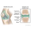
Our Health Library information does not replace the advice of a doctor. Please be advised that this information is made available to assist our patients to learn more about their health. Our providers may not see and/or treat all topics found herein. An arthrogram is a test using X-rays to obtain a series of pictures of a joint after a contrast material (such as a dye, water, air, or a combination of these) has been injected into the joint. This allows your doctor to see the soft tissue structures of your joint, such as tendons, ligaments, muscles, cartilage, and your joint capsule. These structures are not seen on a plain X-ray without contrast material. A special type of X-ray, called fluoroscopy, is used to take pictures of the joint. An arthrogram is used to check a joint to find out what is causing your symptoms or problem with your joint. An arthrogram may be more useful than a regular X-ray because it shows the surface of soft tissues lining the joint as well as the joint bones. A regular X-ray only shows the bones of the joint. This test can be done on your hip, knee, ankle, shoulder, elbow, wrist, or jaw (temporomandibular joint). Other tests, such as magnetic resonance imaging (MRI) and computed tomography (CT), give different information about a joint. They may be used with an arthrogram or when an arthrogram does not give a clear picture of the joint. An arthrogram is used to find the cause of symptoms in your joint. Symptoms may include pain, swelling, or abnormal movement of your joint. It may also be done to see if you can be helped with surgery, such as arthroscopy. An arthrogram can: Tell your doctor before your test if you: The test usually takes about 30 to 60 minutes. You will feel a prick and sting when the anesthetic is given. You may feel tingling, pressure, pain, or fullness in your joint as the dye is put in. The X-ray table may feel hard and the room may be cool. You may have some mild pain, tenderness, and swelling in your joint after the test. Ice packs and nonprescription pain relievers, used as the package directs, may help you feel more comfortable. You may also hear a grating, clicking, or cracking sound when you move your joint. This is normal and goes away in about 24 hours. If you have ongoing pain, tenderness, or swelling of the joint, tell your doctor. You can have a few problems from an arthrogram, such as: There is always a slight risk of damage to cells or tissue from being exposed to any radiation, including the low levels of radiation used for this test. But the risk of damage from the X-rays is usually very low compared with the potential benefits of the test. The radiologist may discuss the initial results with you after reviewing all the pictures. A detailed report will be available to your doctor in a few days. Normal: Abnormal: After seeing the condition of your joint area, your doctor may recommend further treatment with medicine, physical therapy, or surgery. Current as of: March 26, 2025 Author: Ignite Healthwise, LLC Staff Current as of: March 26, 2025 Author: Ignite Healthwise, LLC Staff Clinical Review Board This information does not replace the advice of a doctor. Ignite Healthwise, LLC disclaims any warranty or liability for your use of this information. Your use of this information means that you agree to the Terms of Use and Privacy Policy. Learn how we develop our content. To learn more about Ignite Healthwise, LLC, visit webmdignite.com. © 2024-2026 Ignite Healthwise, LLC.Arthrogram (Joint X-Ray)
Test Overview
Why It Is Done
How To Prepare
How It Is Done
How long the test takes
How It Feels
Risks
Results
Related Information
Credits
Clinical Review Board
All Ignite Healthwise, LLC education is reviewed by a team that includes physicians, nurses, advanced practitioners, registered dieticians, and other healthcare professionals.
All Ignite Healthwise, LLC education is reviewed by a team that includes physicians, nurses, advanced practitioners, registered dieticians, and other healthcare professionals.

Our Health Library information does not replace the advice of a doctor. Please be advised that this information is made available to assist our patients to learn more about their health. Our providers may not see and/or treat all topics found herein. Current as of: March 26, 2025 Author: Ignite Healthwise, LLC Staff Clinical Review Board This information does not replace the advice of a doctor. Ignite Healthwise, LLC disclaims any warranty or liability for your use of this information. Your use of this information means that you agree to the Terms of Use and Privacy Policy. Learn how we develop our content. To learn more about Ignite Healthwise, LLC, visit webmdignite.com. © 2024-2026 Ignite Healthwise, LLC.Arthrogram (Joint X-Ray)
All Ignite Healthwise, LLC education is reviewed by a team that includes physicians, nurses, advanced practitioners, registered dieticians, and other healthcare professionals.